Research Article 
 Creative Commons, CC-BY
Amputation Retractors
*Corresponding author: Fatih Canşah Barışhan, Girne Military Hospital, Turkish Republic of Northern Cyprus, Turkey.
Received: March 13, 2023; Published: April 11, 2023
DOI: 10.34297/AJBSR.2023.17.002479
Abstract
flap survival has increased with the development of anesthetics, surgical facilities, improved techniques, and with the discovery of
antibiotics. In addition, the quality of life for amputation patients has improved significantly with the development of prosthetics.
Today, amputation surgeries can frequently occur due to diabetes and peripheral vascular disease and delay in treatment can be fatal.
During surgery for vasculopathies, retractors are needed to shorten the surgical time for less damage to soft tissues during bone
incision, to protect the vessels of the flap and to make bone incision easier and faster. In this study, 7 different designs of amputation
retractors were compared to the Percy retractor, which is considered the reference design, in terms of physical properties, usage and
marketing. Until the First World War, retractors based on guillotine-like flap-prioritized incisions were used and allowed for more
rapid surgeries. The development of antibiotics led to widespread use of amputation surgery flap applications, the development of
surgical techniques and external surgical instruments. However, with the scientific advances after the Second World War, retractors
were designed to offer both bone incision and flap incision prioritized surgery. In our study, we aimed to determine the development
of amputation retractors in the 19th century and presented suggestions for the design of new retractors to the literature. For new
retractors to be developed, the basic features recommended were those that:
Protect the vessels of the flap while retracting the flap.
Do not to interfere with the surgeon during bone incision.
Offer ease of use with one person.
Can be restabilized.
Do not damage surgical sterile gloves.
Have a large surface area.
Hemostatic.
Keywords: Amputation flap retractor, Retractor for amputation, Amputation instruments, Instruments for amputation surgery, History of amputation retractor, Evolution of amputation retractors, Historical development of amputation retractors
Abbreviations: MESS: Mangled Extremity Severity Score
Introduction
In Spain, France, and New Mexico, there are cave drawings of people with amputations dating back some 36,000 years [1]. In many cultures, amputation was used as a sanction for a judicial punishment. In the Mesopotamian Babylonian culture, the first cases of elective amputation were performed in 1795-1750 BC with the Hammurabi Code, when the tongue was amputated for bad words to the father, one foot for laziness, two arms for rebellion, and one hand for theft [2]. Hippocrates, around 400 BC, recommended amputation for the treatment of gangrene. Hippocrates argued that the best method of removing ischemic tissue is the area closest to the necrotic border, which has lost its vitality and completely lost its sensation. He mentioned the use of cautery and vascular ligature to control bleeding. He also emphasized the need for cauterization, tourniquets, surgical drains, surgical cleaning and antisepsis with wine and vinegar [3]. Albucasis had views that expressed as far back as 1000 years ago and focused on how, “Sometimes the extremities become gangrenous... you must cut off that limb as far as the disease has spread, so that the patient may escape death or greater affliction, greater than the loss of the limb” [4]. Since the 14th century of the Middle Ages, surgical innovations have been many technical developments with the effect of European wars, and with the emergence of firearms, “gunpowder” has been used in the battlefield in amputation surgery for antisepsis and hemostatic purposes [5]. With the advancement in surgical technique and the identification of deaths due to infection and inadequate bleeding control, surgical requirements in this area have increased and Paré proposed the use of tourniquet placement proximal to the amputation site to control bleeding. The rationale was threefold and included appropriate vessel ligation, induction of distal numbness, and facilitation of bone movement. Paré mentioned “phantom pain” for the first time with the use of tourniquets, arguing that phantom pain is less with the use of tourniquets [6]. With the discovery of Sulfuric Ether in 1846, surgeries were initiated and performed more safely and paved the way for the development of many surgical instruments [7]. During the European civil wars and World War I, antimicrobial applications were still limited to topical applications, dressings, and removal of necrotic infected tissues. With the discovery of penicillin by Alexander Fleming in 1928, the development of antibiotics and the availability of sulfonamides in 1938, systemic applications became routine protocols [8]. These advances led to accelerated development of surgical techniques and instruments, and wound healing and infection control can be controlled with systemic applications before and after surgery. The 2nd World War period showed significant differences in the approach to amputation surgeries. With the increased use of weapons of mass destruction, limb loss increased, and the importance of bleeding control increased. National armies included “self-attaching tourniquets” among the standard essential military equipment given to soldiers. With the developments in postoperative rehabilitation techniques and globalized market and marketing policies, prosthesis and orthosis technologies were developed. This has enabled the production of prostheses with high stump compliance at normal stepping speed. For a similar reason, products used in surgeries entered routine use in many amputations’ surgery sets and are now widely preferred and used by surgeons.
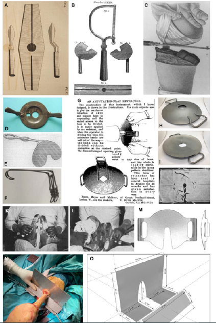
Nowadays, amputations, if unavoidable, are frequently performed in cases of necessity following ischemic and diabetic gangrene. In amputation surgery, retractors are needed as in other surgical interventions in order not to disrupt flap blood supply and to make bone transactions easily during the protection of soft tissues. The first known amputation retractor was described by Gooch in 1767 and in his own words; “In Pl. 8. is the figure of a retraction made of firm strong leather, which I invented and first used in 1739, and, if rightly applied and managed, I am convinced by repeated trials, will effectually answer the purpose.” (Figure 1A) [9]. A drawing of a similar retractor, modified and described by Benjamin Bell in 1796, was described as a c-shaped retractor with one handle made of metal and a rectangular shaped retractor made of leather with a round hole in the center (Figure 1B) [10]. In 1773, William Bromfield, a surgeon from London, recommended pulling the muscles upward during bone incision using a skin compress with a slit in the midline (Figure 1C) [11]. In 1799, Pierre-François PERCY, the first person to develop a triage system in the Napoleonic wars, developed a new retractor for soft tissue packing, consisting of two identical parts articulated by a hinge (Figure 1F) [12]. Even today, the retractor of amputation sets, presented as the “Percy amputation retractor” with a removable handle, is the same as in 18th century models. The retractor, which is shaped like a plate of about 20 cm, has two openings in the center. The smaller one is square, the larger one is circular shaped and 4 cm in diameter. It has handles on both sides, and its single screw-operated arm could wrap around the bone like a guillotine and offered flap incisionprioritized surgery. Cooper, on the other hand, designed a kidneyshaped retractor in 1907 and offered the possibility of single-handed use, but the use of this retractor decreased with the preference of the posterior flap (Figure 1E) [13]. Another amputation retractor used during the First World War was manufactured by Dr. S. J. Streight [14] in 1917 and consisted of L-shaped wire bent like a grid, with one handle (Figure 1F) [15]. In 1918, Harris used his own design circular, double-grip, bone-adjustable retractor for amputation (Figure 1D) [16]. The 1946 “Down Bros and Mayer & Phelps Ltd. A stainless” retractor, in which two kidney-shaped concave semi-circles can move over each other in a way that can be adjusted according to the bone, is exhibited at health museum in south Australia (Figures 1J and 1K) [17]. This retractor is still sold on the market as the “Blake amputation flap retractor” and is still preferred by surgeons in Western Europe today. On the other hand, Lawler stated that his product, which he made in 1946 from a square towel with a round hole in the middle, may also have a place in amputation surgeries from the thigh level (Figure 1L) [18]. Corriero, on the other hand, presented another alternative to literature with her triangular shaped retractor design, which he named Vise amputation retractor [19] in 1957 (Figures 1H and 1I) [20]. Until now, many modifications of the percy retractor have been made. Another remarkable retractor that can be considered original is the double-handled product resembling a cut-out dessert plate called the “Sweet Amputation Retractor”, marketed by Jaret® company through Integra Life Sciences in 2000 (Figure 1M).
Although amputation surgeries were used more frequently during wartime, amputation retractors were mentioned less in the literature during the 1st and 2nd World Wars and different design variants of the Percy retractor were defined. While various retractor designs are needed for surgeons for faster and safer surgeries for amputation surgeries, these designs were a few defined and made, and some of them patented during the insecure times of war. Fewer amputations were made in the post-war years and efforts were made to heal the devastating wounds of the war. It is important to shorten the surgical time in order to reduce the risks related to the operation and anesthesia, especially for today’s amputations performed in patients with advanced age and high comorbidity [21]. For this reason, retractors are still needed in surgery in order to reduce damage to the soft tissues, protect the vessels, and make the bone incision easier and faster. In our study, we aimed to determine the development of amputation retractors in the 19th century and provide insights into this field for the design of new retractors.
Materials and Methods
For systemic analysis, content containing the words “amputation” & “retractor” from 1900 to the present, Google Scholar, Google, PubMed, National Museum Archives of Countries, American-European Patent Institute Records, and related studies were found by searching. For each study, publications/retractors that “describe patented retractors in detail”, “contain retractor drawings or images”, and “designed for amputation surgery” are included. Publications that describe similar retractors, previously defined retractor modifications with the same name, and duplications were excluded from the study. Although not the same retractor, it was included in the study, which was the first defined name for designs that were very similar to the original model, and similar modifications were excluded from the study. For analysis, 3-dimensional structure of retractors, number of handles/handles, adaptability to bone size, moving parts, material construction, direction of use were recorded. Bone retractors are categorically in terms of flap priority, single/two-hand use, assistant needs, number of people and marketing were examined in terms of patentability, manufacturability, and cost. Percy retractor was accepted as the gold standard product due to its current widespread use and modeling diversity, and it was used as a reference design for statistical comparisons. The obtained data were analyzed by grouping, and the properties of patented products were statistically compared with other products.
Results
After the necessary search that met the criteria, a total of 22 models with different designs were found, 6 of them from museum archives, 6 of them from scientific articles, 5 of them from the patent archives of the American patent institute, and 5 of them at the patent pending stage via search engines. Although they contained small details under the name of modification, which were like the Percy retractor, 10 designs were considered as duplications and were excluded from the study. Four of which were not given sufficient technical details were excluded from the design study. Five articles with illustrated design images that will clearly describe the modeled product from scientific articles on amputation retractors were included in our study (Figure 1D, 1E, 1G, 1J, 1K and 1L). Since the sufficient technical details of 2 designs and 1 patented design that were reached from the museum archives were defined, they were included in our study.
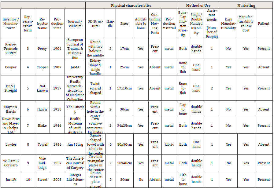
A total of 7 different design amputation retractors were compared with the Percy retractor in terms of physical properties, usage, and marketing. The data are summarized in Table 1.

Table 1:Summary of the results of the comparison with the Percy retractor in terms of physical properties, use and marketing.
All seven retractors were rated as manufacturable at low cost. All the retractors were made by metal except for the towel retractor described by Lawler [16]. All retractors included in the study were capable of surgical use by a single assistant. All but 3 of the retractors were designed to be held with both hands. The long axes of the retractors included in the study were on average 31.6 cm [17-23]. Excluding the Sweet amputation retractor, all patented products were adjustable for bone. Retractors with all moving parts, except Lawler’s fabric retractor, were difficult to manufacture. The Dr. Streight retractor, which can be compressed to a bent gridlike bone and Cooper’s kidney-shaped retractor [8] was designed primarily to cut through bone, while all other retractors included in the study were designed to allow prior flap incision. Mayer & Harris was O-shaped with double handles. Mayer & Harris’ double handle O-shaped retractor and c-shaped double handle dessert plate-like sweet retractor were first designed for flap separation [19]. Except for the kidney-shaped Cooper retractor with a fixed bone socket and the C-shaped sweet retractor, all retractors were able to be adjusted for different diameter bones.
Discussion
The designs of amputation retractors varied during the war years. Surgeons are always looking for devices to shorten the surgical time, and the evolution of retractors and the use of tourniquets have increased over time. However, with the development of angiography and the widespread use of endovascular interventions, the number of ischemic gangrene has decreased, and is in more complex forms [22]. This brings up the necessity of different flap applications and many different under-knee flaps are defined. With the development of microsurgery, grafts, even free flaps can be used today [23]. No matter how advanced the technique is, unfortunately, the use of MESS (mangled extremity severity score) will continue in the future, and the necessity of amputation for ischemic total occluded cases and Wagner grade 4-5 feet will not be eliminated.
For as long as we need it for this treatment method, which has been going on almost since the existence of humanity, in the light of our medical knowledge and possibilities at that time, while dissecting with respect to soft living tissues, we will again take a kind of bone cutter in our hands in order to dominate and separate the hard bone tissue, and at that time we will need to use a retractor. As we mentioned in the historical development, the history of amputation, which started in the form of guillotine surgery in the past, started to allow soft tissue priority incisions that allowed guillotine surgery to be performed. After the concept of posterior flap was introduced for below-knee amputations, the idea of cutting the bone first was tried by the surgeons of that period, Cooper and Straight are among the surgeons who fell into this trend. In this period, which coincided with the First World War, bone incisions were made by placing the designed retractor while preserving the posterior flap vessels behind the tibia with the retractor. After the devastating effects of the war passed, halfring models were designed that would allow both techniques. Harris’ O-shaped closure retractor allowed the bone to be cut by guillotine-like stripping after the flap was separated. According to its contemporaries, it has been evaluated as insufficient because it does not allow the use of the posterior flap. After the war years, the mentioned surgical technique was forgotten, and the retractor design had a stagnation period until the post-World War II years. With the discovery of penicillin following the First World War, the development of antibiotics and their widespread use and the design of amputation retractors was not encountered much in the interwar period. However, during the Second World War, there were significant differences in the approach to amputation surgeries. Despite the developing flap techniques, the retractor technology has not changed, and the use of existing retractors has continued. In the light of this finding and our tertiary center experience on the field offers our design in figure 1N. We named this retractor “Shan amputation retractor” and the technical drawing of the retractor is shown in figure 10.
Conclusion
With the post-war scientific advances, retractors that can offer both bone incision and flap incision surgery have been designed. It is remarkable that the three designs of 1946 and 1957 mentioned in our study are compatible with both techniques. The sweet retractor, which is produced today, is a retractor that primarily allows flap incisions to be made. In this sense, today’s use is more limited than its contemporaries. It is important that these patients with diabetic ischemic disease, whose unfortunate fate is amputation, can be preserved because of the underlying vasculopathy. If flapfirst surgeries are not performed, it is important that the retractors designed today can protect the vessels while offering flap-first opportunities so that the viability of the flap is not affected. In our institutions, where there is often a shortage of extra nurses and assistants, it should be designed in a way that allows surgeons to do it with a single assistant. If it contains moving parts, these must be easily and comfortably flexible to not damage the gloves and their size should not be too large. In our study, we have determined an average value of approximately 30 cm as the average size, it should be in a way that does not tire the weight. In addition, it should be inexpensive and easily sterilizable and suitable for serial surgeries. To traumatize the tissues less, the retraction surface area should be large and flat, hemostatic. We suggest our prototype to current literature.
Acknowledgement
We would like to acknowledge www.makaletercume.com for their outstanding statistical services that were provided for this manuscript..
Conflict of Interests
The authors declare that they have no conflict of interest. The authors did not receive support from any organization for the submitted work. The authors have no relevant financial or nonfinancial interests to disclose.
References
- Magee R (1998) Amputation through the ages: the oldest major surgical operation. Australian and New Zealand Journal of Surgery 68(9): 675-678.
- Horne C F, Johns C H W, King L W (1915) The Code of Hammurabi. Forgotten Books.
- McCollum P T (2005) Limb amputation from aetiology to rehabilitation. R. Ham and L. Cotton. 216 × 138mm. Pp. 229. Illustrated. 1991. London: Chapman and Hall. £95. British Journal of Surgery 79(8): 847-848.
- Iskandar A Z (1975) Albucasis: On Surgery and Instruments. M. S. Spink, G. L. Lewis] 38(2): 440-442.
- Barnes RW, Cox B (2000) Amputations: an illustrated manual.
- Thurston A J (2007) Paré and prosthetics: the early history of artificial limbs. ANZ J Surg 77(12): 1114-1119.
- Hoggard A, Shienbaum R, Mokhtar M, Singh P (2022) Gaseous Anesthetics. In StatPearls.
- Alduina R (2020) Antibiotics and Environment. Antibiotics (Basel) 9(4).
- Gooch B (1767) Cases and Practical Remarks in Surgery with Sketches of Machines Simple in Their Construction Easy in Their Application and of Approved Use (Vol. 2).
- Bell B (1796) Complete course of theoretical and practical surgery at Théophile Barrois the younger. Volume 1.
- Bromfield W (1774) Chirurgische Wahrnehmungen: translated from English and augmented with some additions. Weid-manns Erben und Reich (1st edition ed).
- Bowman H M (1905) Journal des Campagnes du Baron Percy, Chirurgien en Chef de la Grande Armée (1754–1825). Published according to unpublished manuscripts by M. Émile Longin. (Paris: Plon-Nourrit et Cie. 1904. Pp. lxxvii, 537). The American Historical Review 10(2): 417-419.
- Cooper O O (1907) An amputation retractor. Journal of the American Medical Association XLVIII (18): 1506-1506.
- Robertson L B (1918) Further Observations on the Results of Blood Transfusion in War Surgery: With Special Reference to the Results in Primary Hemorrhage. Ann Surg 67(1): 1-13.
- (1917) Research Collection Catalogue. University Health Network - Academy of Medicine Collection.
- Bute Macfie R (1918) An Amputation-Flap Retractor. The Lancet 191(4933): 409.
- (1963) Instrument: Blake Amputation Flap Retractor. Health Museum of South Australia.
- Lawler R H (1946) A towel for use in thigh amputations. Am J Surg 71: 77-79.
- Sweet Amputation Retractor. Retrieved at March 2.
- Corriero W P (1957) A vise-retractor for mid-thigh amputations. Am J Surg 94(4): 683-684.
- Barishan F (2021) Through-Knee Amputation (Knee Disarticulation): A Mini Review of Current Literature. J Diabetic Complications Med 6(5): 155.
- Yolgösteren A, Tok M, Barışhan F C, Kan İ İ (2016) Biyolojik greft ile femoropopliteal baypas uygulanan hastada biyolojik greft anevrizması. TÜRK KALP DAMAR CERRAHİSİ DERNEĞİ KONGRESİ.
- Kümbüloğlu Ö F, Barışhan F C, Özdemir H M, (2021) Two-Stage Flexor Pollicis Longus Tendon Reconstruction Using Pedicled Palmaris Longus Tendon Graft. Muscles Ligaments & Tendons Journal (MLTJ) 11(4): 749-755.


 We use cookies to ensure you get the best experience on our website.